Jul 28 2016
Clemson scientist Jeffrey Anker and four colleagues have been awarded a five-year, $1.57 million grant from the National Institutes of Health to develop a novel imaging technique and dye-based sensor to detect and monitor bacterial infections on implanted medical devices.
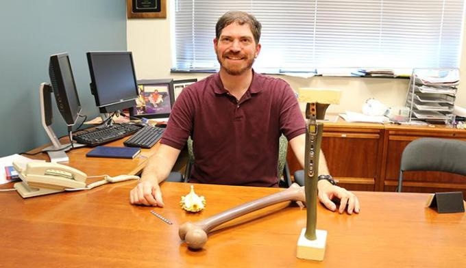
 Principal investigator Jeffrey Anker is an associate professor of chemistry in Clemson University's College of Science. (credit: Jim Melvin / Clemson University)
Approximately one in 25 patients admitted to a hospital in the U.S. will acquire an infection, leading to a reported 99,000 deaths per year. The majority of these hospital-acquired infections involve bacteria growing on implanted medical devices. These devices include metal plates and rods for bone fracture repairs; artificial knees, ankles and hips; prosthetic heart valves, pacemakers and artificial hearts; and urinary and intravascular catheters. Though infections are rare in most implant surgeries, implant-associated infections are difficult and expensive to cure.
"Bacterial colonization of medical implants is a major cause of device failure and often requires device removal coupled with long-term antibiotic treatment," said Anker, associate professor of chemistry in Clemson University's College of Science, with a joint appointment in bioengineering. "However, detection is challenging at early stages when the bacteria are localized to inaccessible regions of the implant. Our research will focus on developing sensors that will coat the implant. Then we'll use X-ray beams to scan the sensors, enabling us to detect and monitor the infection."
With current technologies, there is no effective way to monitor these kinds of infections, either at early stages or during antibiotic treatment when bacteria are not found in the blood. Doctors need a method to monitor the resistant bacteria localized at the implant surface to treat implant infections at early stages and to determine if infections are eradicated.
"If the patient has an infection, the antibiotics will treat the symptoms. But some bacteria often remain on the surface of the implant in the form of a biofilm, which is similar to plaque on your teeth, and the biofilm is resistant to treatment," said Anker, who has spent much of his career studying chemical and biophysical sensors. "But the bacteria that lie dormant within the biofilm's protective environment will periodically sprout and start another infection. At this point, the implant typically needs to be tediously cleansed during a surgical procedure called debridement. If the biofilm isn't fully mature, this is sometimes effective. But if the biofilm has been long established, then debridement usually isn't good enough. The implant will then need to be removed and the remaining infection treated with antibiotics before a new implant is inserted."
Anker's research is designed to find ways to detect implant infections early when they are much easier to cure. With funding from the NIH grant, the team will aspire to develop and improve a dye-based film that will coat implants and serve as an acid sensor. In the human body, most bacteria generate local acidic environments, similar to the process that causes tooth decay. Anker's system will use focused X-ray beams to see through human tissue and strike the coating of the implant in a precise series of layers that will combine to create a computerized image. If an infection is present, the colors revealed in the image will indicate its location and severity.
"What we're attempting to do is quite challenging," Anker said. "We're trying to put a sensor on a plate that will be able to reside in a human body for a reasonable period of time in order to monitor changes in local acidity that will detect infection. Bacteria produce a lot of acids. A human's immune system also produces acids. So if low pH is detected on the surface of an implant, it will be reasonable to assume that the implant is infected. But our research will also delve more into these aspects to determine their validity."
Dr. Tom Pace, professor of orthopedic surgery at the Greenville Health System Department of Orthopedic Surgery and Division Chief of Clinical Sciences at University of South Carolina School of Medicine Greenville, said that implant and orthopedic surgeries are frequently performed and are "highly successful and predictable treatments for millions of patients annually."
"With an aging U.S. population, the expectations are that the next two decades will see a five- to six-fold increase in orthopedic trauma and arthritis implant usage," said Pace, who is one of four co-investigators. "Infections remain infrequent, but they still present significant patient care and cost challenges to the health care system. The Clemson research can help with better treatment and cure rates when these complications do occur, and they can potentially represent cost savings to the community while improving the quality of patient care. That's a win-win for all involved."
Anker is the principal investigator of the project. Listed here are the co-investigators and two laboratory assistants:
- Dr. Caleb Behrend is an adjunct professor at Clemson as well as a practicing orthopedic surgeon and educator at Virginia Tech Carilion School of Medicine and Research Institute in Roanoke, Virginia. Behrend has extensive experience in sensor design, synthesis and materials design. He will assist with research-related surgeries performed on cadaver and animal models.
- John DesJardins is an associate professor at Clemson and the Hambright Leadership Associate Professor in Bioengineering. He has worked as a biomedical research engineer for more than 20 years. After the chemistry of the sensor operation is determined, DesJardins' lab will help to design, fabricate, test and validate the implants under realistic conditions.
- Dr. Tom Pace is an adjunct professor of bioengineering at Clemson, in addition to his responsibilities at GHS. He will join Behrend in assisting with research-related surgeries performed on cadaver and animal models.
- T.R. Jeremy Tzeng is an associate professor at Clemson and the graduate program coordinator for microbiology in the biological sciences department. Tzeng has extensive experience in pathogen-host interactions. He will assist in developing the infection models and determining the biocompatibility of the sensors.
- Unaiza Uzair is a Fulbright scholar from Pakistan who will assist with the development of the sensors in Anker's lab at Clemson.
- Donald Benza is a master's electrical engineering student at Clemson who will develop and improve equipment and software for the scanning system in Anker's lab.
"We want to be able to diagnose infections before they become catastrophic problems," Behrend said. "I'm excited to be a part of this team and am pleased that the National Institutes of Health has recognized the value of our research."