Jul 3 2014
Optical imaging methods are rapidly becoming essential tools in biomedical science because they’re noninvasive, fast, cost-efficient and pose no health risks since they don't use ionizing radiation. These methods could become even more valuable if researchers could find a way for optical light to penetrate all the way through the body's tissues.
With today’s technology, even passing through a fraction of an inch of skin is enough to scatter the light and scramble the image.
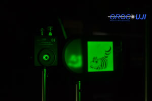
 The researchers placed an image of the Cheshire Cat in front of a scattering layer that completely hid it, then used the image they were able to retrieve from photodetected values recorded by the photodiode to demonstrate that disorder-assisted imaging can indeed recover an object's fine details -- even using a sensor without spatial resolution. (Photo credit: Jesús Lancis/Jaume I University.)
Now a team of researchers from Spain’s Jaume I University (UJI) and the University of València has developed a single-pixel optical system based on compressive sensing that can overcome the fundamental limitations imposed by this scattering. The work was published today in The Optical Society’s (OSA) open-access journal Optics Express.
“In the diagnostic realm within the past few years, we’ve witnessed the way optical imaging has helped clinicians detect and evaluate suspicious lesions," said Jesús Lancis, the paper’s co-author and a researcher in the Photonics Research Group at UJI. "The elephant in the room, however, is the question of the short penetration depth of light within tissue compared to ultrasound or x-ray technologies. Current knowledge is insufficient for early detection of small lesions located deeper than a millimeter beneath the surface of the mucosa."
"Our goal is to see deeper inside tissue,” he added.
To achieve this, the team used an off-the-shelf digital micromirror array from a commercial video projector to create a set of microstructured light patterns that are sequentially superimposed onto a sample. They then measure the transmitted energy with a photodetector that can sense the presence or absence of light, but has no spatial resolution. Then they apply a signal processing technique called compressive sensing, which is used to compress large data files as they are measured. This allows them to reconstruct the image.
One of the most surprising aspects of the team’s work is that they use essentially a single-pixel sensor to capture the images. While most people think that more pixels result in better image quality, there are some cases where this isn't true, Lancis said. In low-light imaging, for instance, it's better to integrate all available light into a single sensor. If the light is split into millions of pixels, each sensor receives a tiny fraction of light, creating noise and destroying the image.
“Something similar happens when you try to transmit images through scattering media,” Lancis said. “When we use a conventional digital camera to get an image, we only see the familiar noisy pattern known as ‘speckle.’ In compressive imaging, since we aren’t using pixelated sensors, it should be less sensitive to light scrambling and enable transmission of images through scattering.”
Also notable, the team’s technique could operate through dynamic scattering. “Most scattering media of interest, like biological tissue, are dynamic in the sense that the scatter centers continuously change their positions with time -- meaning that the speckle patterns are ‘in motion.’ This is ideal for some applications because monitoring the changes of the speckle can reveal information about the sample, but the drawback is that it’s a major nuisance to transmit or get images,” Lancis pointed out. “Our technique, however, requires no calibration of the medium, and its fluctuations during the sensing stage don’t limit imaging ability.”
What’s ahead for the team? “Our next goal is to break the barriers of light penetration depth inside a scattering medium with the state-of-the-art megapixel-programmable spatial light modulators used in consumer electronics,” Lancis says. To do this, they’ll need to demonstrate that their technique works even when the sample is embedded inside the tissue.
Paper: “Image transmission through dynamic scattering media by single-pixel photodetection,” E. Tajahuerce et al., Optics Express, Vol. 22, Issue 14, pp. 16945-16955 (2014).